Imagine two photographs of the same face, taken several months apart. In the second, the skin looks smoother. A treatment has worked, perhaps. Or the light has moved. Or the chin has tilted. A photograph can tell the truth and still give you entirely the wrong impression. The trouble begins when someone asks it to serve as evidence.
Canfield Scientific has made that trouble its business. It builds cameras, lighting systems, image software, and the services that help clinicians and researchers capture comparable pictures. Its unglamorous obsession is consistency. Before anyone can measure a change in skin, someone has to stop the photography from changing.
That is a surprisingly useful position to occupy. A dermatologist needs to locate a lesion again. A plastic surgeon needs to explain a possible alteration in shape. A drug developer needs to document outcomes across study sites. Each customer has a different question, but all three need pictures that can survive a second look.
- The problem: changes in pose and light can masquerade as changes in skin.
- The product: connected imaging hardware, software, and repeatable capture procedures.
- The buyers: medical practices and sponsors of clinical studies.
- The lesson: control the observation before measuring the outcome.
02The real invention is the repeat visit
Founded by Doug Canfield, the privately held company sits at the intersection of medical photography, dermatology equipment, and clinical research. Its expertise spans optics, 2D and 3D capture, image management, and analysis. Think of a photographic studio whose operating instructions have become part of the product.
Consider IntelliStudio. Software controls zoom, lighting, and camera orientation; templates guide the patient’s pose. The motorized lift takes care of positioning. MatchPose overlays an earlier image onto the live view so that the operator can align the follow-up. The baseline photograph becomes an instruction for the next photograph.
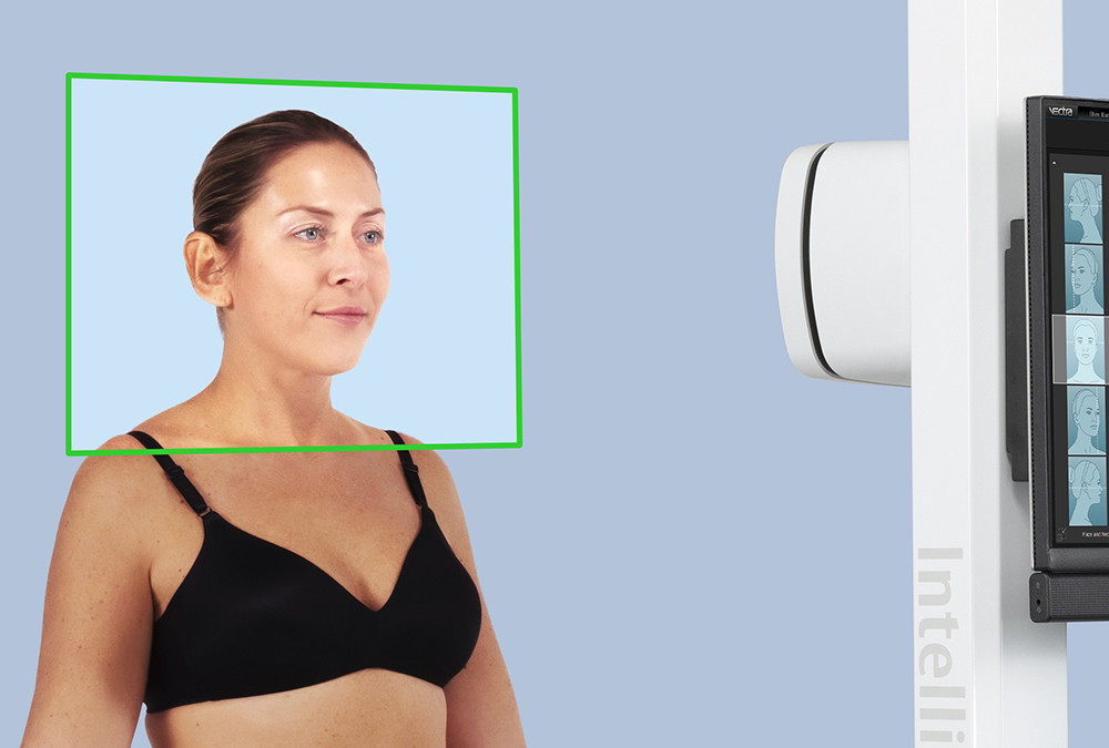
Canfield also sells a turntable, IntelliStage. This is a pleasingly literal solution to a clinical photography problem: sometimes the easiest way to obtain another view is to turn the person. Its published capacity is 150 kilograms, and rotation can be controlled by remote, flash trigger, or Bluetooth with the VECTRA H2.
The transferable lesson is simple: standardize the conditions before comparing the results. Specify the pose, the lighting, the capture settings, and the follow-up procedure. A shiny analysis screen cannot repair a comparison that was unfair before the shutter fired.
03A face, a scalp, an entire body
The product range follows the scale of the question. VISIA examines facial skin using standard, cross-polarized, and ultraviolet lighting. It analyzes features including spots, wrinkles, texture, pores, and red areas. Different illumination brings different features into view; the ordinary photograph is only one account of the face.
On March 25, 2026, Canfield launched VISIA 3D, bringing VECTRA-derived 3D tools and VISIA skin analysis into one consultation platform. The launch describes one-click capture of the front and both sides of the face, plus a full-face 3D image. Measurement, contouring, and simulation tools let a practitioner discuss shape as well as complexion.
The portable VECTRA H2 and stationary VECTRA XT address face, breast, and body imaging for aesthetic consultations. Mirror software handles medical image management. These tools give a patient and practitioner a shared visual object to discuss. A simulation illustrates a proposed change; it should be read as a consultation aid, rather than a promised surgical result.
Then there is HairMetrix, which performs automated hair analysis without clipping the hair. It combines close-up imaging, baseline comparisons, and patient reports. Anyone who has tried to describe hair loss with the words “a little more than last time” can appreciate the attraction of a measurement.
04Forty-six pairs of eyes
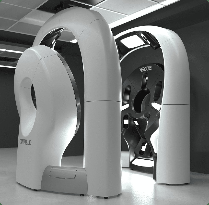
At the other end of the scale is the VECTRA WB360. Its 46 stereo-vision pods create a 3D whole-body image in a single capture, recording exposed skin and body shape. The machine looks extravagant until you consider the bookkeeping problem it addresses: which mark, on which part of the body, changed since the previous visit?
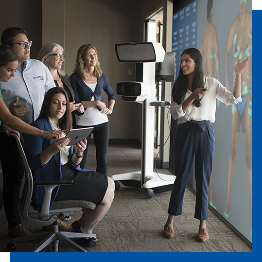
DermaGraphix links close-up dermoscopy images to the body map and gives lesions unique identifiers. Overview and detailed images can be reviewed together. Markerless tracking and guided follow-up help clinicians return to the same places. The image becomes an address system.
This is where Canfield’s work becomes more consequential than a polished before-and-after display. Longitudinal skin surveillance depends on continuity. A clinician needs the history of a particular lesion, rather than a folder of nearly identical elbows. Organizing that history is a substantial part of the product’s practical value.
On September 29, 2026, Canfield announced EU Medical Device Regulation CE marking for two AI tools: DEXI, Class IIb, for supporting skin-cancer assessment, and Lesion Visualizer, Class IIa, for detecting lesion change. The announcement specifies trained, experienced clinicians as users. Outside the EU, both tools remain for research use only. Geography and intended use matter here as much as the algorithm.
“The future of dermatology involves AI and the clinician working together.”
Brian D’Alessandro, Ph.D.
Canfield Principal AI Scientist · September 2026
05The business behind the consultation room
The less visible half of Canfield is its clinical-services operation. Pharmaceutical, biotechnology, medical-device, and cosmetics businesses hire it to support study imaging. Work runs from protocol design and site equipment to image review, analysis, data management, and presentation. Canfield reports more than 4,200 clinical studies and more than 11 million images processed. Those are company-reported totals, not a count of successful treatments.
Canfield-reported cumulative totals. Volume measures experience, not treatment efficacy.
Canfield Capture software is configured around a study protocol. It controls camera settings, collects subject information electronically, and sends images to the clinical-services website for review. Researchers can compare images and track study status. The process makes repeatability a shared obligation across sites rather than a photographer’s private knack.
- 01DefineProtocol, views, lighting
- 02CaptureGuided, repeatable images
- 03ReviewQuality checks and queries
- 04AnalyzeMeasurements and assessments
Its analysis work includes measurements such as surface area, volume, and skin movement. Clinical data services include reconciliation and third-party integration. For a sponsor, the purchase is therefore larger than a camera: it is a way to manage the photographic part of a trial with equipment, people, and procedures that fit together.
The business model follows this division. Practices buy imaging hardware and software, with support and training through Canfield Care. Sponsors commission clinical services. The combination gives Canfield practical contact with both everyday consultations and research requirements - a useful basis for deciding what its next instrument should do.
06The invoice includes a workflow
Canfield’s product pages invite prospective buyers to request a quote. A sensible purchasing exercise includes computers, storage, networking, staff training, and ongoing support alongside the imaging system. For WB360, Canfield lists up to 3 GB of data per patient visit and a minimum 4 TB server drive. The impressive photograph has an equally real storage bill.
Operational fit matters. Published WB360 specifications require wired workstation connectivity and do not support cloud storage for live data, although cloud backup is allowed. Those requirements should be checked against a practice’s infrastructure before ordering. A clinic expecting the entire system to behave like a casual phone app would be buying the wrong workflow.
Adoption can begin with skepticism. In a testimonial published on the VECTRA XT page, surgeon Simeon Wall Jr. recalls partners doubting its usefulness and cost before becoming advocates. This is a vendor-selected account, not an independent return-on-investment calculation. It illustrates the adoption question: will the team actually use the extra information during consultations?
Alternatives exist. FotoFinder offers total-body mapping and dermoscopy; QuantifiCare’s LifeViz systems serve 3D imaging. Canfield’s distinctive pitch is the breadth of its connected hardware, software, and clinical services. That breadth matters most when a buyer needs several parts to work together. For a narrower task, the comparison should begin with that task’s requirements.
07A company that buys closer views
Canfield’s expansion has also been optical. It acquired Germany’s VISIOMED in 2018, adding dermoscopy expertise and establishing a European center for high-magnification imaging. In 2021 it acquired Italy’s Medici Medical, creating an Italian sales, support, and logistics operation and launching the Dreamoscopy education program.
Its stated approach to development is a small-team process of planning, prototyping, and iteration. The company describes commercial and clinical staff working across projects, and its clinical-services page says 30 percent of staff work in research and development. These are useful clues to how it wants to operate, rather than a substitute for employees’ own accounts of working there.
There is a tension running through this portfolio. Better pictures can educate patients and help practices sell treatments; standardized images can also support research and medical assessment. The pictures may look equally persuasive while doing different jobs. Canfield is most interesting when it makes the job explicit.
For the reader, the lesson travels well beyond skin. If you want to know whether something improved, make the next observation comparable to the first. Canfield has built an extensive business around the details of that instruction. The humble repeat photograph turns out to require rather a lot of engineering.
See the instruments at work
Explore Canfield’s website, its product range, clinical services, and news and product updates.
Watch product demonstrations on Canfield’s YouTube channel or the WB360 capture demonstration. Follow the company on LinkedIn, Instagram, Facebook, and X.